In the fibular hemimelia is the congenital absence or underdevelopment of the fibula (fibula medical term). The disease is also known as a longitudinal fibular defect. It can occur either in isolation or in combination with malformations of the thigh bone, malformations of the feet or with a shortening of the entire lower leg bone.
What is a fibular hemimelia?
According to LAWFAQS.NET, fibular hemimelia is a disease that is very rare. It only occurs in three out of 100,000 newborns. Fibular hemimelia affects men twice as often as women. In two thirds of all sick people, only one leg is affected by the malformation.
Fibular hemimelia is more common on the right leg than on the left leg. The congenital, complete absence of a shin bone, which is known as the tibial hemimelia, is even less common. The fibular hemimelia is characterized by a congenital and longitudinal defect and has a wide range of deformities.
Causes
Potential causes of fibular hemimelia have not yet been adequately researched to be able to provide reliable information about them. However, the search for the causes of the disease has been the subject of medical research for a long time. Fibular hemimelia has a familial accumulation, which leads to the conclusion that it is a congenital disease with hereditary causes.
The malformation could have its origin in the disturbance of a critical period in the development of the limbs in the embryo. This lies in the period between the fourth and seventh week of pregnancy. In addition, viral infections, vascular dysgenesis, trauma and various environmental influences are discussed as potential causes. An autosomal dominant inheritance was inferred from a familial occurrence of fibular hemimelia.
Symptoms, ailments & signs
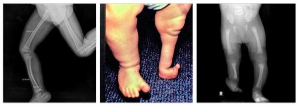
The symptoms and complaints associated with fibular hemimelia can be varied. As a functional symptom of the disease, the shortening of the affected leg is in the foreground, which increases in the course of the growth phase. In most cases, the foot is also affected by the disease, often in what is known as a kinked and equinus foot position.
Only medial rays of the affected foot are applied. In addition, parts of the rear foot are sometimes missing. In numerous cases, the entire lower leg is underdeveloped or hypoplastic. In addition, there is often a femoral hypoplasia in connection with the fibular hemimelia. A so-called ball joint deformity in the ankle joint with impaired joint mechanics is often associated with fibular hemimelia.
Other malformations that often occur in combination with fibular hemimelia can be, for example, a proximal femur defect, malformations of the toes or a craniosynostosis. Fibular hemimelia can also be associated with skeletal dysplasia.
The bone affected by the disease has only a low growth potential, so that the deformity increases with age. In rare cases, fibular hemimelia is associated with other malformations that are outside the skeletal system, such as heart defects, thrombocytopenia, kidney dysplasia or malformations of the eyes. However, intellectual deficits rarely occur in the context of fibular hemimelia.
Diagnosis
Various examination-technical methods are available for the diagnosis of fibular hemimelia. An existing malformation of the embryo can already be detected in the womb by means of an ultrasound examination. After birth, the warping and shortening of the affected bones are evident. Carrying out X-ray examinations provides information about the severity of the malformation and additional changes in the bones.
In any case, the diagnosis must be made both clinically and radiologically. Differential diagnoses must also be carried out in order to be able to differentiate fibular hemimelia from various other diseases with sometimes similar symptoms. This is particularly relevant in light of the fact that each disease requires individual therapy despite similar complaints. In particular, the presence of thalidomide embryopathy, amniotic ligament syndrome and various skeletal dysplasias with asymmetrical involvement of the lower limbs should be checked.
Complications
This disease leads to severe malformations and deformities in the area of the feet and thighs. In most cases, these deformities also restrict movement and reduce aesthetics. Many patients also experience inferiority complexes and reduced self-esteem.
These symptoms can sometimes lead to depression. In addition to the malformations of the skeleton and the feet, complaints of the heart and kidneys can also occur. In the worst case, these symptoms can be fatal if kidney failure or heart failure occurs. In some cases, developmental disorders can also occur that lead to decreased intelligence.
The treatment itself does not lead to any further complications and can alleviate the symptoms of the disease relatively well. Above all, surgical interventions are used to bring the legs to the same length. An amputation may be necessary. If there are further problems with coordination or movement, therapeutic measures are used. If the organs are spared from damage, life expectancy remains unchanged.
When should you go to the doctor?
If anomalies and malformations of the skeletal system are noticed during the birth of a child, a medical examination should be initiated immediately. Inpatient births are accompanied by an obstetrician team who independently check the infant’s state of health. If the birth takes place without an obstetrician, a doctor must be consulted immediately afterwards. Inconsistencies in the bone structure of the calf or legs are considered a matter of concern.
A doctor should be consulted as soon as palpable irregularities on the feet, thighs or lower legs can be perceived. If the toe formation is visually noticeable, a check-up should be initiated. If there are clearly recognizable deformities during the growth and development process of the child, a doctor should be consulted. If the child’s growth is significantly delayed compared to that of their peers, especially in the area of the legs or feet, a check-up should be carried out. If joint problems and irregularities occur, the observations should be discussed with a doctor.
If the child experiences emotional or mental problems in the course of life due to the optical anomalies, a doctor or therapist should be consulted. In the case of aggressive behavior, reduced self-esteem or a strong tendency to withdraw, it is advisable to provide the child with therapeutic help. If serious psychological problems arise in the form of an anxiety disorder or depression, the child needs professional help.
Treatment & Therapy
For the treatment of fibular hemimelia, various measures can be considered, which are based on the individual case. In any case, treatment should be started as early as possible and, if possible, take place in a pediatric orthopedic center. The spectrum of possible treatment methods ranges from orthoses and prostheses to so-called conversion osteotomies.
In more severe cases, surgical leg lengthening or even amputation of the affected leg must be considered. The weaker the fibular hemimelia, the more it can be expected that the leg will remain in good working order. In general, the therapy takes place in connection with a multidisciplinary approach, whereby genetic counselors and pediatric orthopedic surgeons work together.
The aim of orthopedic treatment is to compensate for the leg length discrepancy and, in bilateral cases, to correct the asymmetrical short stature. If the fibular hemimelia is only weakly pronounced and has a slight discrepancy in leg length, therapy using orthopedic shoes and appropriate insoles is also used to compensate for the difference in length.
Outlook & forecast
The prospect of recovery from fibular hemimelia is very good. If the condition is treated surgically or with medication immediately after the birth, it can be relieved relatively quickly. However, the child needs support in the form of physiotherapy.
In some cases, for example if the fibular hemimelia significantly restricts mobility, an outpatient nurse must also be called in to help the child with everyday tasks. Basically, the prognosis is good if the patient receives the necessary treatment and later comprehensive support.
The prognosis is worse if the condition is not recognized in time or the therapy does not show the desired effect. Then permanent restrictions of the ability to move can remain. In addition, it can lead to paresthesia, nerve damage and other complications that significantly reduce the quality of life.
The life expectancy is not restricted by the fibular hemimelia. As a result of the disease, the risk of circulatory disorders, thromboses and other complications that can be life-threatening increases. After an initial examination and the start of therapy, the doctor can make a prognosis and suggest further measures.
Prevention
Since fibular hemimelia is most likely a hereditary disease, there are no known effective methods of preventing the malformation. It is all the more important to carry out specialist examinations if there are signs of the disease in order to positively influence the further course of the fibular hemimelia.
Aftercare
Since this disease is a congenital disease, no causal, but only purely symptomatic therapy can be carried out. A complete cure is also not possible. If the patient wishes to have children, genetic counseling and testing can also be carried out so that the disease is not passed on to the descendants.
The earlier the disease is recognized, the better the further course will usually be. The treatment itself is primarily carried out by wearing prostheses. These should always be used if they can completely alleviate the symptoms. In severe cases, however, surgical interventions or even an amputation are necessary to completely limit this disease.
After such an operation, the person concerned usually has to rest and in any case take care of his body. In doing so, strenuous activities or stressful activities should be avoided in order not to slow down the healing process. Furthermore, in many cases, psychological support for the person affected is also useful.
This can also be carried out by relatives or friends, whereby contact with other patients of this disease can also be useful. The life expectancy of the person affected remains unaffected by this disease.
You can do that yourself
If fibular hemimelia has been detected in a newborn, treatment should be started as early as possible. In consultation with the responsible doctor in the maternity hospital, contact must be made promptly with a specialized clinic in order to enable the child to receive the best possible therapy. Also orthopedists and specialists for specific symptoms should be promptly turned on, for only so smooth therapy is possible.
Parents who have psychological problems as a result of the child’s illness are advised to consult a therapist. Participation in a self-help group may also be useful. The affected child may also need psychological support later in life.
Depending on the extent of the fibular hemimelia, the child may need crutches or a prosthesis. After an amputation, rest is indicated. The child will have to spend the first few weeks in the clinic and needs extensive support from the parents at home.
In some cases, an outpatient care service must be called in. Since dealing with an affected child is a significant burden, all available support and support options should be used. Regardless of the severity of the malformation, a close medical check-up of the patient is necessary.